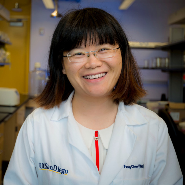
Dr. Fang Chen
Graduate student researcher
University of California, San Diego
Dr. Fang Chen completed her Ph.D. in the Program of Materials Science and Engineering at UCSD, and will become a postdoctoral researcher at the Byers Eye Institute and Spencer Vision Research Center at Stanford University. During her Ph.D., Dr. Chen worked on the development of ultrasound-based contrast agents and theranostic nanoparticles for regenerative medicines in treating heart diseases. Dr. Chen has published more than 16 peer-reviewed papers and contributed more than 14 presentations for professional conferences. She also has 2 patent disclosures and 2 chapters. Dr. Chen also serves as a reviewer for Acta Biomaterialia and Journal of Biomaterials Application. To recognize her accomplishments in research and mentorship, Dr. Chen has been awarded the Chancellor’s Research Excellence Scholarships (CRES) and the MATS Dissertation Year Fellowship.
Inorganic Nanoparticles Increasing Stem Cell Contrast and Therapeutic Efficacy in Myocardial Infarction Treatment
Cardiovascular diseases are a leading cause of death. Although the existing therapies can lower early mortality rates and reduce the risk of further heart attacks, patients can suffer from existing damage for their rest of life due to the very limited regeneration capacity of heart. Therefore, many efforts have been made to regenerate and repair cardiac tissue via stem cells. However, stem cell therapy in cardiovascular diseases treatment is challenged by mis-injection, poor survival, and low retention of cells. Solutions to these challenges can be multimodal imaging contrast agents, sustained or controlled drug delivery, and magnetic manipulation of cells, all of which bring attention to nanotechnology. In this dissertation, I will demonstrate how nanotechnology can benefit stem cell therapy with inorganic silicon-based nanomaterials and mesenchymal stem cells. Mesenchymal stem cells (MSCs) are used in this dissertation because they are particularly promising due to their abundance, potent proliferation, and multi-lineage differentiation capacity.
First, the role of hybrid nanoparticles in multi-modal imaging and theranostic nanomedicine and an introduction to medical imaging modalities and therapeutic methods are reviewed. The second chapter describes a novel discoid silica nanoparticle, which offered the most enhanced ultrasound signal of MSCs compared to three other classical silica structures including Stober silica, mesoporous silica, and mesocellular foam silica. The third chapter first introduce the biocompatibility of silicon carbide nanomaterials. Silicon carbide nanowires inhibited proliferation and differentiation of MSCs while silicon carbide nanoparticles showed no impact on the MSCs’ viability and functions. In the second part, the potential of silicon carbide nanoparticles in stem cell long-term imaging/tracking via photoacoustic and photoluminescence imaging is shown. The fourth chapter studies the drug adsorption behavior of organosilica nanoparticles with intrinsic amine groups.
At last, a biocompatible multi-functional silica-iron oxide nanoparticle is designed. This nanoparticle increases MRI and ultrasound contrast, cell viability, and retention of MSCs. In vivo studies show that labeling stem cells with this nanoparticle increase the stem cell therapy efficacy in murine animals injured by a ligation/reperfusion surgery.